

Surgeons and all doctors are in constant need of different medical imaging techniques. X-ray, NMR, Ultrasonic, and other methods all provide valuable information from the inside of a patient. We have examined the possibilities that infrared imaging offers to the doctors, concentrating in TRAM patients.
IR imaging is based on the subject's own heat production, and the method is thus noninvasive. A standard IR camera can be used, typically with spatial resolution of 1x1mm at one meter distance, and about 0.05 centigrade heat resolution. The output can immediately be seen or the data can be recorded and analyzed later. The idea is that IR camera could detect the blood vessels that come to the surface from the body's heat sources - inner organs and muscles. The surgeons can use the information in the planning of the operation. The local temperatures of the transplant can easily be tracked after the operation, and the heat distribution tells if something is going wrong in it.
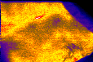 Beginning - the abdominal area intact
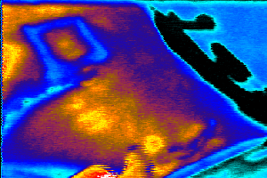 Random phase, flap is half-cut
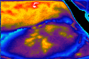 Still attached, almost dissected
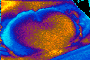 Dead phase - the flap is off
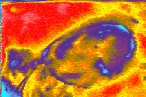 The new breast constructed from the flap